Lincoff’s Rules: Localising the Retinal Break
Read the configuration of subretinal fluid, predict where the causative break is most likely to lie, and know where to search first.
Watch the lecture for the animated retinal-detachment configurations, then use this companion note for structured revision and clinical application.
Why localising the break matters
A rhegmatogenous retinal detachment (RRD) develops when liquefied vitreous passes through a full-thickness retinal break into the subretinal space, separating the neurosensory retina from the retinal pigment epithelium.
Identifying the causative break — and looking carefully for additional breaks — is therefore a central part of evaluating an RRD and planning treatment.
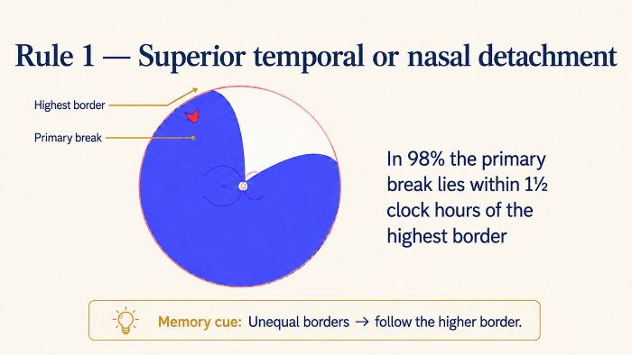
Lincoff and Gieser showed that the configuration of subretinal fluid is not random. Its spread is influenced by the location of the break, anatomical boundaries and gravity, so the shape of the detachment can help predict where the primary break is most likely to lie. [1]
The rules are therefore best used as a search guide: they tell you where to look first, not where to stop.

Where are retinal breaks commonly found?
Approximate proportions of retinal-break location in rhegmatogenous retinal detachment are: [3]
The superotemporal quadrant is therefore the commonest location. However, the entire retinal circumference must still be examined carefully.
When more than one break is present, an additional break may lie within 90° of the first break in approximately 50% of eyes. [3]
Read the detachment before looking for the break
Before applying an individual Lincoff rule, first describe the configuration of the retinal detachment. Five observations usually narrow the area in which the primary break should be sought.
-
1Is the detachment mainly superior or inferior?
-
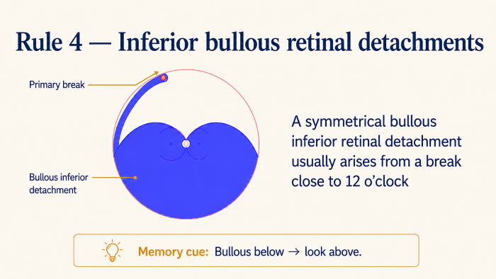
2Is it shallow or bullous?
-
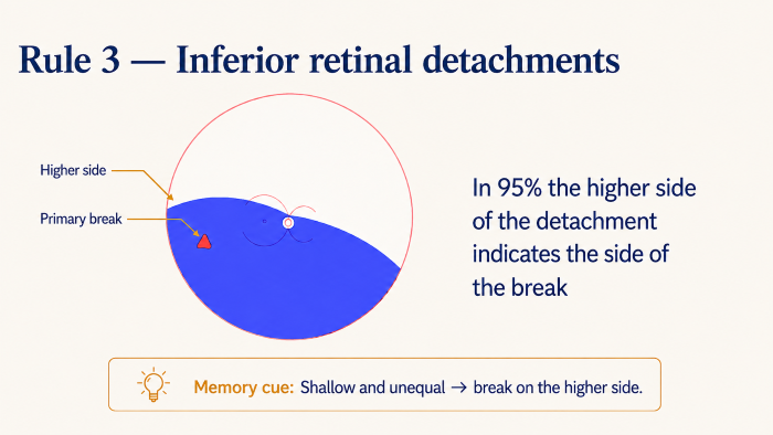
3Are the nasal and temporal fluid levels symmetrical or asymmetrical?
-
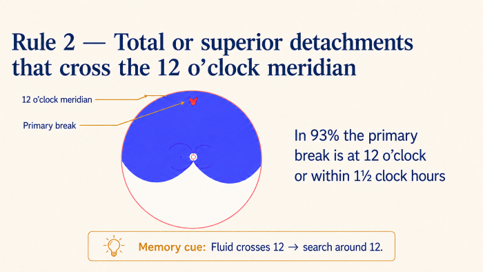
4Does the subretinal fluid cross the 12-o’clock meridian?
-
5Which is the highest border of the detachment?
These five observations form the framework for interpreting the four classic Lincoff patterns.
Rule 2 — Superior or total detachment crossing 12 o’clock
When a superior or total retinal detachment crosses the 12-o’clock meridian, the primary retinal break is most likely to lie at or close to 12 o’clock, within the superior search zone extending approximately 1½ clock hours to either side. This relationship was reported in approximately 93% of cases. [1] [2]
Supplementary patterns
These additional configurations extend the same principles of subretinal-fluid spread and break localisation. [1] [3] [4]
Symmetrical shallow inferior detachment
When a shallow inferior detachment has approximately equal nasal and temporal fluid levels, the retinal break is usually located inferiorly near 6 o’clock.
Near-total detachment with an inferior attached wedge
If most of the retina is detached but an inferior wedge remains attached, with subretinal fluid communicating superiorly across the 12-o’clock meridian, search the superior retina around 12 o’clock.
Subtotal detachment with a superior attached wedge
When a superior wedge remains attached and subretinal fluid does not freely cross the superior vertical meridian, search the peripheral retina near the highest border of the detachment.
Bullous subtotal detachment
If a subtotal detachment is markedly bullous, the causative break is likely to be superior. Begin the search near the highest superior extent of the fluid.
A practical localisation approach
Rather than memorising every configuration in isolation, assess the detachment in the same sequence each time.
Is it bullous?
A markedly bullous inferior detachment should immediately raise suspicion of a superior retinal break.
Is it superior or inferior?
In a superior detachment, assess the highest border and whether fluid crosses 12 o’clock. In an inferior detachment, decide whether it is shallow or bullous and whether the borders are equal.
Are the borders unequal?
If one side rises higher than the other, search toward the higher border.
Does fluid cross 12 o’clock?
If superior subretinal fluid crosses the vertical meridian, concentrate the search around 12 o’clock.
Pseudophakic RRD
In pseudophakic rhegmatogenous retinal detachment, retinal breaks may be small, peripheral or multiple and can be more difficult to identify. Lincoff’s rules can help direct the initial search, but a meticulous examination of the peripheral retina remains essential. [6]
Rapid revision
- • First decide whether the detachment is shallow or bullous.
- • Unequal borders direct the search toward the higher border.
- • If superior subretinal fluid crosses 12 o’clock, search around 12 o’clock.
- • A shallow inferior detachment usually points toward an inferior break.
- • A symmetrical shallow inferior detachment suggests a break near 6 o’clock.
- • A bullous inferior detachment should prompt a careful search superiorly.
Related Videos
Retinal Detachment: Types, Risk Factors, Pathophysiology, Symptoms and Signs
Review the clinical framework of retinal detachment before applying break-localisation rules.
Principles of Retinal Detachment Repair
Continue from break localisation to the principles used to plan retinal-detachment repair.
References
-
1.
Lincoff H, Gieser R. Finding the retinal hole. Arch Ophthalmol. 1971;85(5):565–569. doi:10.1001/archopht.1971.00990050567007
-
2.
Sultan ZN, Agorogiannis EI, Iannetta D, Steel D, Sandinha T. Rhegmatogenous retinal detachment: a review of current practice in diagnosis and management. BMJ Open Ophthalmol. 2020;5(1):e000474. doi:10.1136/bmjophth-2020-000474
-
3.
Turgut B. Remembering the Modified Lincoff’s rules for practical identification of retinal break in rhegmatogenous retinal detachment. Adv Ophthalmol Vis Syst. 2018;8(5):267–268. doi:10.15406/aovs.2018.08.00318
-
4.
Yoshida I, Shiba T, Hori Y, Maeno T. Relationships between retinal break locations and the shapes of the detachments. Clin Ophthalmol. 2018;12:2213–2222. doi:10.2147/OPTH.S177594
-
5.
Xiong J, Tran T, Waldstein SM, Fung AT. A review of rhegmatogenous retinal detachment: past, present and future. Wien Med Wochenschr. 2025;175(7–8):186–202. doi:10.1007/s10354-025-01085-9
-
6.
Gupta D, Ching J, Tornambe PE. Clinically undetected retinal breaks causing retinal detachment: a review of options for management. Surv Ophthalmol. 2018;63(4):579–588. doi:10.1016/j.survophthal.2017.08.002