Diabetic Retinopathy: Why the Retina Leaks and Becomes Ischaemic
Understand how persistent hyperglycaemia injures the retinal neurovascular unit and produces the two central processes of diabetic retinopathy: vascular leakage and capillary non-perfusion.
Watch the Lecture
Watch the lecture first, or use the companion note below to revise how hyperglycaemia leads to retinal leakage, non-perfusion and neovascularisation.
The Path from Hyperglycaemia to Retinopathy
Capillary-wall injury and barrier breakdown → microaneurysms, retinal haemorrhages, hard exudation and diabetic macular oedema
Capillary closure and dropout → retinal ischaemia, VEGF release and neovascularisation
Duration Creates Cumulative Risk
The risk of diabetic retinopathy rises with the duration of diabetes and cumulative glycaemic exposure.
Important risk modifiers
- Long-term glycaemic control
- Hypertension
- Diabetic kidney disease
- Dyslipidaemia
- Pregnancy in patients with pre-existing diabetes
Better glycaemic and blood-pressure control reduces the development and progression of diabetic retinopathy. [1] [6] [7]
Classic epidemiological teaching suggested that after approximately 20 years of diabetes, nearly 99% of people with type 1 diabetes and more than 60% of those with type 2 diabetes had developed some degree of retinopathy.
These are historical estimates from older cohorts; the younger-onset Wisconsin cohort reported retinopathy in approximately 97.5% after 15 years or more. [2] [3]
Contemporary estimates are lower and more variable. One type 1 diabetes cohort reported retinopathy in approximately 54% at 20 years, while a modern type 2 diabetes cohort reported approximately 53% prevalence at 20–25 years.
Rates vary between populations, treatment eras and screening methods. [4] [5]
Hyperglycaemia Creates Metabolic Stress
Persistent hyperglycaemia activates several interacting biochemical pathways that converge on oxidative stress, inflammation and retinal neurovascular dysfunction. [8] [9] [10]
Polyol Pathway
Excess intracellular glucose is converted to sorbitol by aldose reductase, using NADPH.
NADPH is also required to regenerate reduced glutathione, an important intracellular antioxidant. Excessive NADPH consumption therefore reduces glutathione-dependent antioxidant capacity.
Sorbitol is then converted to fructose by sorbitol dehydrogenase, using NAD⁺ and producing NADH.
Sorbitol oxidation therefore increases the intracellular NADH:NAD⁺ ratio, producing redox imbalance and potentially increasing reactive oxygen species generation.
Advanced Glycation
Glycation is the non-enzymatic attachment of reducing sugars to proteins, lipids or nucleic acids.
With persistent hyperglycaemia, these early glycation products undergo further modification to form advanced glycation end products (AGEs).
AGEs alter proteins and extracellular matrix and can activate the receptor for advanced glycation end products (RAGE).
AGE–RAGE signalling promotes:
- Oxidative stress
- Inflammation
- Endothelial dysfunction
- Abnormal extracellular-matrix turnover
- Leukocyte adhesion
Other Hyperglycaemic Pathways
Hyperglycaemia also activates:
- Protein kinase C
- The hexosamine biosynthetic pathway
- Mitochondrial production of reactive oxygen species
These pathways further disturb vascular permeability, blood-flow regulation, inflammatory signalling and extracellular-matrix metabolism.
The hexosamine pathway is distinct from the pentose-phosphate pathway. [8] [9]
From Metabolic Stress to Capillary Injury
Metabolic, oxidative and inflammatory stress damages the principal components of the retinal capillary wall.
Pericyte Loss
Pericytes provide structural support, regulate capillary tone and help maintain endothelial stability. Their loss weakens the capillary wall and contributes to microaneurysm formation.
Endothelial Dysfunction
Endothelial cells regulate retinal blood flow, vascular permeability and leukocyte interaction. Their tight junctions form the inner blood–retinal barrier; dysfunction therefore contributes to both vascular leakage and capillary non-perfusion.
Basement-Membrane Thickening
Abnormal extracellular-matrix turnover disrupts endothelial–pericyte interaction and impairs capillary function.
Together, these changes lead to the two major pathways of diabetic retinopathy:
Vascular Leakage
Leakage in diabetic retinopathy reflects two related but distinct consequences of retinal capillary injury: capillary-wall fragility and breakdown of the inner blood–retinal barrier.
Capillary-Wall Damage
Loss of pericyte and endothelial support weakens the retinal capillary wall and makes it structurally unstable.
Barrier Breakdown
Dysfunction of endothelial tight junctions weakens the inner blood–retinal barrier and increases vascular permeability.
Clinical Manifestations
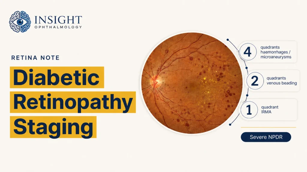
Focal outpouchings arising from structurally weakened retinal capillaries.
Blood escapes from damaged or fragile retinal microvessels.
Lipid-rich residues remain after leakage of plasma constituents into the retina.
Retinal thickening caused by blood–retinal barrier breakdown and vascular leakage involving the macula.
Capillary Non-Perfusion and Retinal Ischaemia
Endothelial injury, leukocyte adhesion and progressive capillary degeneration can cause retinal capillaries to close and disappear from the functioning circulation.
Cotton-Wool Spots
Cotton-wool spots are focal accumulations of axoplasmic material caused by interrupted axoplasmic transport in ischaemic regions of the retinal nerve-fibre layer.
Venous Beading
Venous beading describes irregular variation in retinal venous calibre and reflects significant disturbance of the retinal circulation.
IRMA
Intraretinal microvascular abnormalities are abnormal intraretinal vascular channels found beside areas of capillary non-perfusion.
From Ischaemia to Neovascularisation
When capillary non-perfusion becomes extensive, the ischaemic retina increases production of angiogenic mediators—most importantly vascular endothelial growth factor (VEGF).
NVD
Neovascularisation of the disc refers to new vessels arising at or near the optic disc.
NVE
Neovascularisation elsewhere refers to new vessels arising away from the optic disc on the retinal surface.
Proliferative Disease
The presence of NVD or NVE defines proliferative diabetic retinopathy.
Why New Vessels Threaten Vision
Neovascular tissue is structurally fragile and may bleed into the pre-retinal space or vitreous cavity.
New vessels grow with accompanying fibrovascular tissue along the retinal and posterior hyaloid surfaces.
Contraction of fibrovascular tissue can pull on the retina and produce tractional retinal detachment.
Do Not Confuse: IRMA and Neovascularisation
Both are associated with retinal ischaemia, but they represent different vascular responses and have different implications for disease staging.
| Feature | IRMA | Neovascularisation |
|---|---|---|
| Full term | Intraretinal microvascular abnormalities | New vessels at the disc or elsewhere |
| Location | Remain within the retina | Grow on the retinal or optic-disc surface |
| Mechanism | Remodelling of existing intraretinal vascular channels beside areas of capillary non-perfusion | Angiogenic growth of new vessels in response to retinal ischaemia and increased VEGF |
| Fluorescein leakage | Usually limited or absent | Typically prominent |
| Disease significance | Prominent IRMA is a feature of severe non-proliferative diabetic retinopathy; the presence of any IRMA does not automatically establish severe NPDR | NVD or NVE defines proliferative diabetic retinopathy |
From Mechanism to Clinical Sign
The retinal signs of diabetic retinopathy become easier to remember when each is linked to its dominant underlying mechanism.
| Dominant process | Underlying event | Common manifestations |
|---|---|---|
| Capillary-wall instability | Pericyte loss and endothelial dysfunction weaken the retinal capillary wall | Microaneurysms and retinal haemorrhages |
| Barrier breakdown | Endothelial tight-junction dysfunction increases vascular permeability | Hard exudates, retinal thickening and diabetic macular oedema |
| Capillary non-perfusion | Capillary degeneration, closure and dropout produce retinal ischaemia | Cotton-wool spots, venous beading and IRMA |
| Angiogenic response | Ischaemic retina increases VEGF production | NVD, NVE and proliferative diabetic retinopathy |
Clinical Summary
The pathogenesis of diabetic retinopathy can be consolidated into five connected principles.
Cumulative exposure determines risk
Longer diabetes duration and sustained hyperglycaemic exposure increase the likelihood of retinal injury, although modern outcomes are modified by glycaemic and systemic risk-factor control.
Hyperglycaemia activates interacting injury pathways
Polyol-pathway activity, advanced glycation, protein kinase C, hexosamine-pathway activity and mitochondrial oxidative stress converge on inflammation and retinal neurovascular dysfunction.
Retinal capillary injury has two major outcomes
Pericyte loss, endothelial dysfunction, abnormal extracellular matrix and leukostasis lead to vascular leakage or capillary non-perfusion.
Leakage produces oedema and exudation
Capillary-wall instability produces microaneurysms and retinal haemorrhages, while blood–retinal barrier breakdown produces hard exudates, retinal thickening and diabetic macular oedema.
Non-perfusion produces ischaemia and proliferation
Capillary closure produces retinal ischaemia, which increases VEGF production and may progress to NVD, NVE and proliferative diabetic retinopathy.
Related Videos
Continue revising diabetic retinopathy through the complete Insight Ophthalmology video playlist.
References
- Lim JI, et al. Diabetic Retinopathy Preferred Practice Pattern®. Ophthalmology. 2025;132(4):P75–P162. doi:10.1016/j.ophtha.2024.12.020
- Fong DS, Aiello L, Gardner TW, et al. Diabetic retinopathy. Diabetes Care. 2003;26 Suppl 1:S99–S102. doi:10.2337/diacare.26.2007.S99
- Klein R, Klein BEK, Moss SE, Davis MD, DeMets DL. The Wisconsin Epidemiologic Study of Diabetic Retinopathy. II. Prevalence and risk of diabetic retinopathy when age at diagnosis is less than 30 years. Arch Ophthalmol. 1984;102(4):520–526. doi:10.1001/archopht.1984.01040030398010
- Madeira C, Lopes M, Laiginhas R, et al. Changing trends in the prevalence of diabetic retinopathy in type 1 diabetes mellitus from 1990 to 2018: a retrospective study in a Portuguese population. Diabetes Res Clin Pract. 2019;158:107891. doi:10.1016/j.diabres.2019.107891
- Voigt M, Schmidt S, Lehmann T, et al. Prevalence and progression rate of diabetic retinopathy in type 2 diabetes patients in correlation with the duration of diabetes. Exp Clin Endocrinol Diabetes. 2018;126(9):570–576. doi:10.1055/s-0043-120570
- Diabetes Control and Complications Trial Research Group. The effect of intensive treatment of diabetes on the development and progression of long-term complications in insulin-dependent diabetes mellitus. N Engl J Med. 1993;329(14):977–986. doi:10.1056/NEJM199309303291401
- UK Prospective Diabetes Study Group. Tight blood pressure control and risk of macrovascular and microvascular complications in type 2 diabetes: UKPDS 38. BMJ. 1998;317(7160):703–713. doi:10.1136/bmj.317.7160.703
- Brownlee M. Biochemistry and molecular cell biology of diabetic complications. Nature. 2001;414(6865):813–820. doi:10.1038/414813a
- Duh EJ, Sun JK, Stitt AW. Diabetic retinopathy: current understanding, mechanisms, and treatment strategies. JCI Insight. 2017;2(14):e93751. doi:10.1172/jci.insight.93751
- Lechner J, O’Leary OE, Stitt AW. The pathology associated with diabetic retinopathy. Vision Res. 2017;139:7–14. doi:10.1016/j.visres.2017.04.003
- Joussen AM, Murata T, Tsujikawa A, Kirchhof B, Bursell SE, Adamis AP. Leukocyte-mediated endothelial cell injury and death in the diabetic retina. Am J Pathol. 2001;158(1):147–152. doi:10.1016/S0002-9440(10)63952-1
- Aiello LP, Avery RL, Arrigg PG, et al. Vascular endothelial growth factor in ocular fluid of patients with diabetic retinopathy and other retinal disorders. N Engl J Med. 1994;331(22):1480–1487. doi:10.1056/NEJM199412013312203
- Cheung N, Mitchell P, Wong TY. Diabetic retinopathy. Lancet. 2010;376(9735):124–136. doi:10.1016/S0140-6736(09)62124-3