Insight Ophthalmology · Retina Companion Note
Classification of Diabetic Retinopathy
Diabetic retinopathy is staged according to the retinal abnormalities visible on examination. This companion note brings together the practical clinical scale, the examination-important 4–2–1 rule, progression risks and the historical high-risk PDR criteria.
Diabetic macular oedema is assessed separately and may coexist with any retinopathy stage.
Watch the lecture first, then use this companion note to consolidate the clinical stages, the 4–2–1 rule and the progression risks.
The Classification at a Glance
The International Clinical Diabetic Retinopathy Disease Severity Scale follows five practical stages, progressing from no visible retinopathy to proliferative disease. [1]
retinopathy
Very severe NPDR is an ETDRS-derived category defined by two or more components of the 4–2–1 rule. In the simplified international scale, it is included within severe NPDR.
High-risk PDR is a DRS-derived category identifying proliferative disease associated with a particularly high risk of severe visual loss.
Clinical Scale and Study Classifications
Several closely related classification systems appear in diabetic retinopathy teaching. They serve different purposes and should not be treated as interchangeable.
ETDRS grading
The Early Treatment Diabetic Retinopathy Study used a detailed photographic grading system derived from the modified Airlie House classification. Lesions were compared with standard stereoscopic fundus photographs, making the scale highly precise but relatively complex for routine clinical use. [2]
International clinical scale
The International Clinical Diabetic Retinopathy Disease Severity Scale simplified the evidence into five practical clinical stages. [1]
- No apparent retinopathy
- Mild NPDR
- Moderate NPDR
- Severe NPDR
- Proliferative diabetic retinopathy
DRS high-risk criteria
The Diabetic Retinopathy Study identified high-risk characteristics within PDR. These criteria were designed to recognise eyes with a particularly high risk of severe visual loss and a clear benefit from prompt panretinal photocoagulation. [4]
Clinical Scale and Study Classifications
Several closely related classification systems appear in diabetic retinopathy teaching. They serve different purposes and should not be treated as interchangeable.
ETDRS grading
The Early Treatment Diabetic Retinopathy Study used a detailed photographic grading system derived from the modified Airlie House classification. Lesions were compared with standard stereoscopic fundus photographs, making the scale highly precise but relatively complex for routine clinical use. [2]
International clinical scale
The International Clinical Diabetic Retinopathy Disease Severity Scale simplified the evidence into five practical clinical stages. [1]
- No apparent retinopathy
- Mild NPDR
- Moderate NPDR
- Severe NPDR
- Proliferative diabetic retinopathy
DRS high-risk criteria
The Diabetic Retinopathy Study identified high-risk characteristics within PDR. These criteria were designed to recognise eyes with a particularly high risk of severe visual loss and a clear benefit from prompt panretinal photocoagulation. [4]
From No Apparent Retinopathy to Moderate NPDR
The early stages are distinguished by the number and type of visible retinal lesions, while neovascularisation remains absent. [1]
No apparent retinopathy
No visible diabetic retinal abnormalities.
The patient has diabetes, but no clinically apparent diabetic retinopathy is detected on examination. [1]
Mild NPDR
Microaneurysms only.
Microaneurysms are the only visible diabetic-retinopathy lesion. No additional findings are present to justify a higher stage, and there is no neovascularisation. [1]
Moderate NPDR
More than microaneurysms alone, but less than severe NPDR.
The retina may show one or more of the following:
- Intraretinal dot-and-blot haemorrhages
- Hard exudates
- Cotton-wool spots
- Venous abnormalities
- Limited intraretinal microvascular abnormalities
The eye does not fulfil any component of the severe-NPDR 4–2–1 rule and has no signs of proliferative disease. [1]
Severe NPDR: The 4–2–1 Rule
Severe NPDR is diagnosed when any one of the following criteria is present, provided there is no retinal or optic-disc neovascularisation. [1]
Intraretinal haemorrhages
More than 20 intraretinal haemorrhages in each of all four retinal quadrants.
Venous beading
Definite venous beading in two or more retinal quadrants.
IRMA
Prominent intraretinal microvascular abnormalities in one or more retinal quadrants.
What are IRMA?
Intraretinal microvascular abnormalities are abnormal vascular channels that develop within areas of capillary non-perfusion. They remain within the retina and are not themselves neovascularisation. [2]
Severe and Very Severe NPDR
The distinction depends on how many components of the 4–2–1 rule are present. Neovascularisation remains absent in both categories. [2] [3]
| Feature | Severe NPDR | Very Severe NPDR |
|---|---|---|
| 4–2–1 rule | Any one criterion | Any two or more criteria |
| Neovascularisation | Absent | Absent |
| Detailed ETDRS grading | Levels 53A–53D | Level 53E |
| Simplified international scale | Classified as severe NPDR | Included within severe NPDR |
| Progression risk | High Approximately 50% progress to any PDR and 15–17% to high-risk PDR within 1 year. | Very high Approximately 75% progress to any PDR and 45% to high-risk PDR within 1 year. |
The progression percentages are historical ETDRS-derived natural-history estimates. [3]
Why the Stage Matters
The risk of proliferative disease rises sharply once severe NPDR is reached. The figures below are historical ETDRS-derived estimates and show why severe and very severe NPDR should not be treated as equivalent. [3]
Severe NPDR
-
≈50%
at 1 year Progression to any PDR -
≈17%
at 1 year Progression to high-risk PDR -
≈71%
at 3 years Progression to any PDR -
≈44%
at 3 years Progression to high-risk PDR
Very Severe NPDR
-
≈75%
at 1 year Progression to any PDR -
≈45%
at 1 year Progression to high-risk PDR
Proliferative Diabetic Retinopathy
The transition from NPDR to PDR occurs when retinal or optic-disc neovascularisation develops. [1] [5]
PDR is diagnosed when definite retinal or disc neovascularisation is present, with or without associated preretinal or vitreous haemorrhage.
Defining findings
- Neovascularisation of the disc
- Neovascularisation elsewhere on the retina
- Associated preretinal haemorrhage
- Associated vitreous haemorrhage
Why the vessels bleed
The new vessels are fragile and lack the normal structural stability of mature retinal vessels. They may bleed into the potential space in front of the retina or into the vitreous cavity.
Fibrovascular contraction
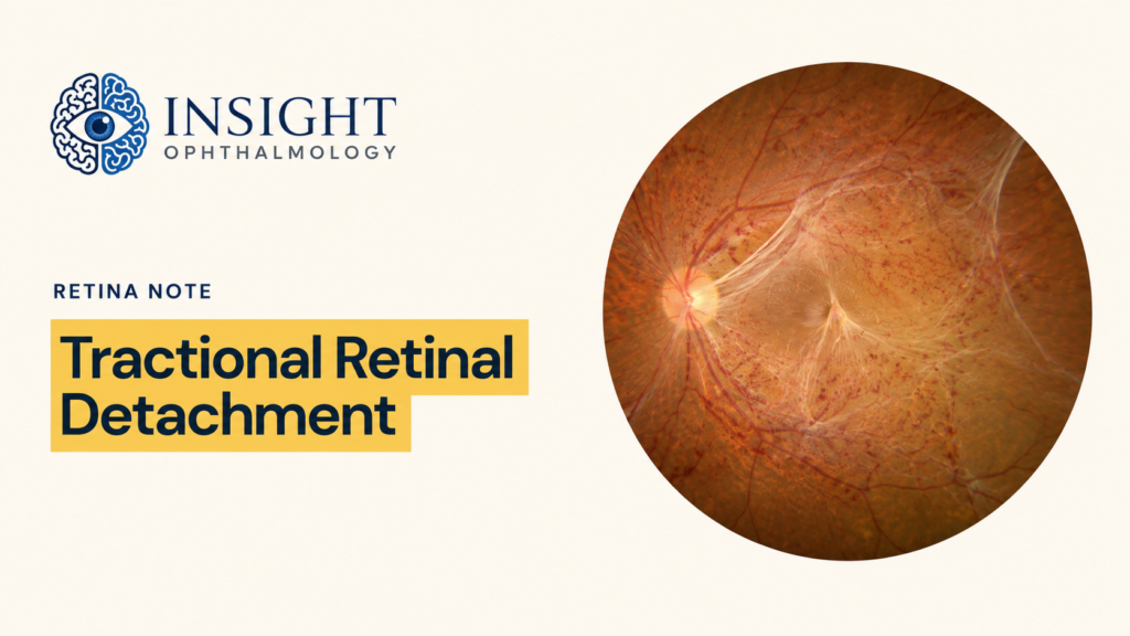
Neovascular tissue is often accompanied by a fibrous component. Contraction of this fibrovascular tissue can exert traction on the retina and produce a tractional retinal detachment.
Where Neovascularisation Appears
The site of the new vessels determines whether they are described as NVD or NVE. Either finding establishes proliferative diabetic retinopathy. [1]
Neovascularisation of the disc
NVDNew vessels located on the optic disc or within approximately one disc diameter of the disc.
The extent of NVD and the presence of preretinal or vitreous haemorrhage are important when applying the historical DRS high-risk criteria.
Neovascularisation elsewhere
NVENew vessels arising on the retinal surface away from the disc region.
NVE may be accompanied by preretinal or vitreous haemorrhage and may develop a fibrovascular component capable of producing retinal traction.
Anterior-segment neovascularisation
New vessels may also develop on the iris or within the anterior chamber angle in eyes with extensive retinal ischaemia.
This finding carries a risk of neovascular glaucoma and indicates severe ocular ischaemia. [5]
High-Risk PDR: The DRS Criteria
The Diabetic Retinopathy Study identified particular patterns of neovascularisation associated with a high risk of severe visual loss. These historical criteria remain important for examinations and for understanding the evidence behind panretinal photocoagulation. [4]
Substantial NVD
Neovascularisation of the disc at least as extensive as DRS standard photograph 10A—approximately one-quarter to one-third disc area.
Preretinal or vitreous haemorrhage is not required when NVD is this extensive.
Any NVD with haemorrhage
NVD of any extent associated with preretinal haemorrhage or vitreous haemorrhage.
Extensive NVE with haemorrhage
Neovascularisation elsewhere measuring at least approximately one-half disc area, together with preretinal or vitreous haemorrhage.
Why was it called high risk?
These eyes had a particularly high risk of severe visual loss.
In the DRS, severe visual loss meant visual acuity below 5/200 at two consecutive follow-up examinations, four months apart. [4]
Retinopathy Stage and Macular Oedema
Diabetic macular oedema is assessed separately because it may occur at any stage of diabetic retinopathy. [1] [5]
Each eye should therefore be described using two parallel assessments: the retinopathy stage and the presence or absence of diabetic macular oedema.
Two separate axes
- Retinopathy severity describes the overall retinal stage: no apparent DR, NPDR or PDR.
- DME status describes whether retinal thickening is present in the macula and whether the foveal centre is involved.
Centre-involving DME
Retinal thickening involves the foveal centre or central OCT subfield.
Non-centre-involving DME
Retinal thickening is present within the macula but spares the foveal centre.
Classification Summary
Classify the eye according to the most advanced retinal finding present, then document diabetic macular oedema separately. [1] [5]
No apparent retinopathy
No visible diabetic retinal abnormalities.
Mild NPDR
Microaneurysms only.
Moderate NPDR
More than microaneurysms alone, but less than severe NPDR.
Severe NPDR
Any one component of the 4–2–1 rule, with no neovascularisation.
Very severe NPDR
Any two or more components of the 4–2–1 rule, with no neovascularisation. [2]
PDR
Definite NVD or NVE, with or without associated preretinal or vitreous haemorrhage.
High-risk PDR
A historical DRS-defined subgroup of PDR with a particularly high risk of severe visual loss. [4]
Classification Summary
Classify the eye according to the most advanced retinal finding present, then document diabetic macular oedema separately. [1] [5]
No apparent retinopathy
No visible diabetic retinal abnormalities.
Mild NPDR
Microaneurysms only.
Moderate NPDR
More than microaneurysms alone, but less than severe NPDR.
Severe NPDR
Any one component of the 4–2–1 rule, with no neovascularisation.
Very severe NPDR
Any two or more components of the 4–2–1 rule, with no neovascularisation. [2]
PDR
Definite NVD or NVE, with or without associated preretinal or vitreous haemorrhage.
High-risk PDR
A historical DRS-defined subgroup of PDR with a particularly high risk of severe visual loss. [4]
Continue Learning
Use these related lectures to recognise the individual retinal lesions, distinguish IRMA from neovascularisation and assess diabetic macular oedema.
References
Landmark classification studies, progression evidence and current clinical guidance used in this companion note.
-
1.
Wilkinson CP, Ferris FL III, Klein RE, et al; Global Diabetic Retinopathy Project Group. Proposed international clinical diabetic retinopathy and diabetic macular edema disease severity scales. Ophthalmology. 2003;110(9):1677–1682.
doi:10.1016/S0161-6420(03)00475-5 International five-stage clinical scale, simplified 4–2–1 rule and separate assessment of diabetic macular oedema. -
2.
Early Treatment Diabetic Retinopathy Study Research Group. Grading diabetic retinopathy from stereoscopic color fundus photographs—an extension of the modified Airlie House classification: ETDRS Report Number 10. Ophthalmology. 1991;98(5 Suppl):786–806.
doi:10.1016/S0161-6420(13)38012-9 Detailed ETDRS photographic grading, lesion severity and ETDRS levels 53A–53E. -
3.
Early Treatment Diabetic Retinopathy Study Research Group. Fundus photographic risk factors for progression of diabetic retinopathy: ETDRS Report Number 12. Ophthalmology. 1991;98(5 Suppl):823–833.
doi:10.1016/S0161-6420(13)38014-2 Photographic predictors of progression and historical progression risks for severe and very severe NPDR. -
4.
Diabetic Retinopathy Study Research Group. Photocoagulation treatment of proliferative diabetic retinopathy: clinical application of Diabetic Retinopathy Study findings—DRS Report Number 8. Ophthalmology. 1981;88(7):583–600.
doi:10.1016/S0161-6420(81)34978-1 High-risk PDR characteristics, severe visual loss and the landmark benefit of photocoagulation. -
5.
Lim JI, Kim SJ, Bailey ST, et al; American Academy of Ophthalmology Preferred Practice Pattern Retina/Vitreous Committee. Diabetic Retinopathy Preferred Practice Pattern®. Ophthalmology. 2025;132(4):P75–P162.
doi:10.1016/j.ophtha.2024.12.020 Current terminology, OCT assessment and centre-involving versus non-centre-involving diabetic macular oedema.